VASCULAR.EXPERT
mini-invasives
technics

Marc FITOUSSI M.D
Vascular Surgeon.
18-22 Queen Anne Street
W1G 8HU London
+44 20 7034 3326
VASCULAR.EXPERT
Marc FITOUSSI M.D
Vascular Surgeon.
18-22 Queen Anne Street
W1G 8HU London
+44 20 7034 3326
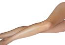
ANATOMY
great saphen vein small saphen vein
NORMAL SYSTEMIC CIRCULATION
The oxygen-rich blood leaves the heart and flows through the arteries, bringing oxygen to every organ, and tissue, all the way to the tip of your hands and your feet. Once the oxygen in the blood is used by these organs and tissues, the blood will then return to the heart, using the veins.
In the legs, the blood should then go up and back to the heart. To help in this process, the veins are equipped with anti-reflux valves, which prevent the blood from going down, under the influence of gravity.
They are found by pair, along the veins, with a decreasing density from the end of the limbs to the body.
The veins are said to be continent when the blood flow is normal, going either up, or from the periphery to the body, and incontinent when the blood flow is going the wrong way. This leads to varicose veins.
VENOUS ECHO-DOPPLER
This is done on a patient standing up, and it is the standard test to detect varicose veins by analyzing:
1 - Deep venous trunks for permeability and continence.
2- Superficial venous trunks : great and small saphenous veins,
ANATOMY
In the lower limbs, there are two venous networks: A deep one, and a superficial one.
It is the superficial one that is responsible for the common varicose veins.
Profound network veins :
-Sural veins:
Peroneal veins, anterior and posterior tibial veins.
Origin: the feet
Ending: ring of the soleus, where they merge to form the popliteal vein
-Popliteal vein :
Origin: the knee, merge with the smaller saphenous, and twin veins.
Ending: ring of the third adductor, the vein is continued by the superficial femoral vein.
-Femoral veins:
The deep and superficial femoral veins merge to become the common femoral vein, which receives the great saphenous vein. It ends at the crural canal.
-The veins coming from the limbs then reach the abdomen, by the iliac veins, which then meet to form what is called the inferior vena cava which brings the blood back to the heart.
The superficial veins :
Located at the surface of the limbs, it accounts for 10% of the blood circulation.
There are two superficial veins per leg:
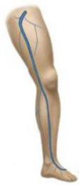
-The internal saphenous vein, also called the great or greater saphenous vein, which starts next to the internal malleolus of the ankle, then ends up in the femoral vein in the groin.
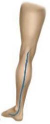
-The external saphenous vein, also called the smaller or small saphenous vein, which begins behind the external malleolus of the ankle, and ends up in the popliteal vein in the trick knee.
There are others accessory superficial veins like:
-The anterior saphenous vein, which travels the anterior part of the thighs, and the external part of the legs
-The posterior saphenous vein,
-Some transverse collaterals which connect internal and external saphenous veins.
The deep and superficial networks can communicate by several ways :
The normal pathway of the blood is from the superficial veins to the deeper ones, which brings the blood back to the heart.
-The internal saphenous vein’s arch, which ends up in the common femoral vein
-The external saphenous vein’s arch, which ends up in the popliteal vein
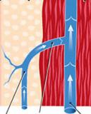
-The perforating veins, storied along the limbs.
The denomination “perforating” is reserved for anastomoses between superficial and deep veins, “perforating” the aponeurosis.
The perforating veins have anti-reflux valves which prevents the flow from going from the wrong way, the right way being from the superficial veins to the deeper ones
VASCULAR.EXPERT
mini-invasives technics
18-22 Queen Anne Street W1G 8HU London
+44 20 7034 3326